
Vascular Malformation and AVM Assessment
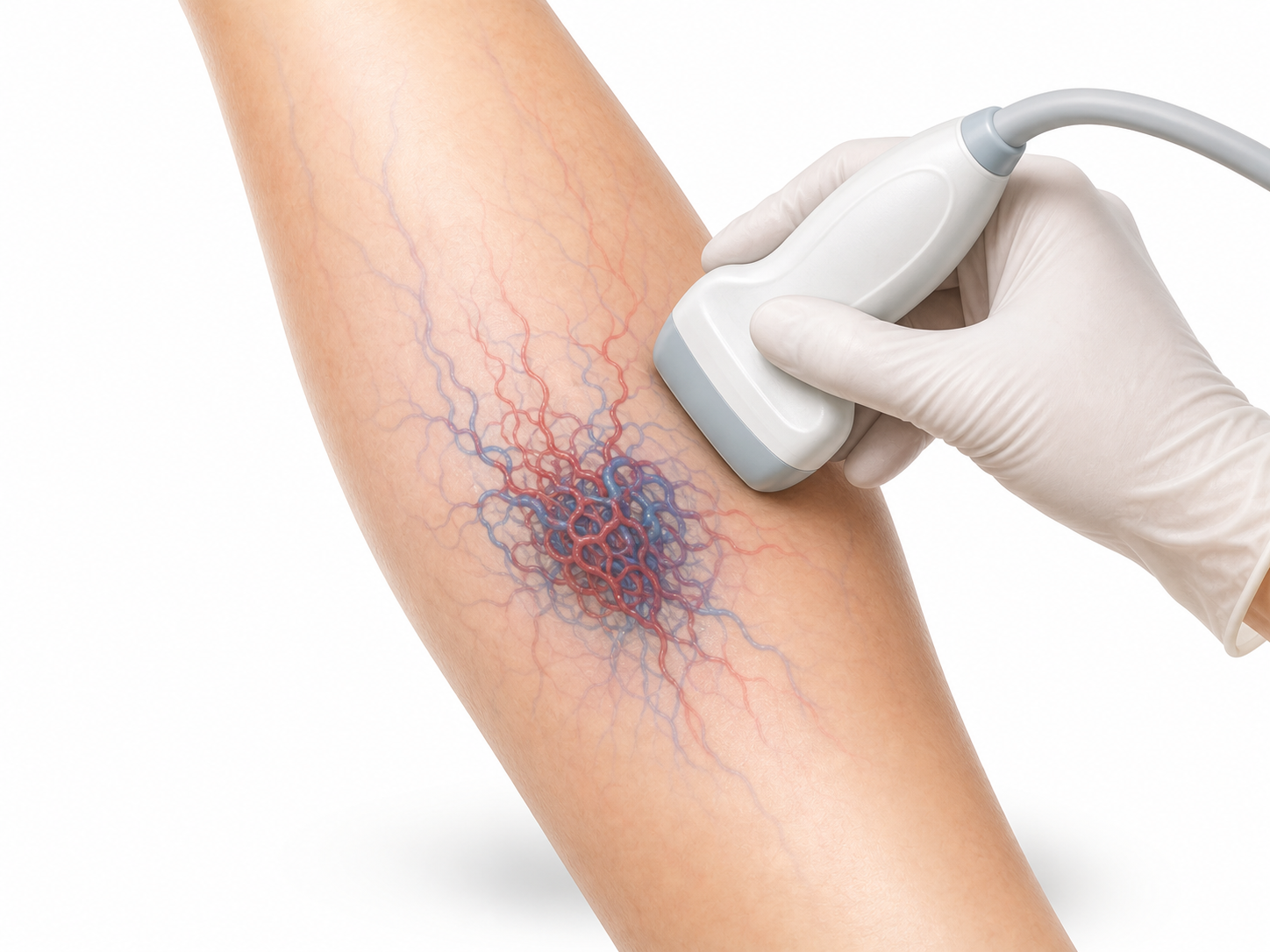
A Doppler mapping visit for selected vascular anomalies, abnormal superficial vessels, suspected AVM, or a mass where flow pattern matters.
Duration
45-60 minutes
Location
Salmiya
Visit Time
45-60 minutes
Preparation
Bring prior MRI, CT, ultrasound, or specialist notes if available.
Clinic
International Hospital, 2nd Floor, Vascular Department
Best for
When the question is vascular
What ultrasound contributes
Ultrasound can show whether a lesion has venous, arterial, or high-flow features and can help document depth, flow direction, and vessels involved.
Why prior imaging is useful
Vascular anomalies often need combined information from clinical exam and imaging. Prior MRI or CT helps focus the Doppler exam and report.
Bleeding, rapid growth, severe pain, skin breakdown, or neurological symptoms need prompt medical review.
For sudden or severe symptoms, call Kuwait emergency services on 112 or go to the nearest emergency department. Do not wait for a WhatsApp reply.
Booking Path
From question to report
Prepare the location, duration, symptoms, and prior imaging for the visit. Do not send them through WhatsApp; wait for a secure channel if requested.
The scan maps flow and visible vessels.
The report supports referral or follow-up planning.
Patient Questions
Practical details before the visit
This information is general and does not replace physician assessment. For severe symptoms, call Kuwait emergency services on 112 or go to the nearest emergency department.
Related Services
You may also need
Pseudoaneurysm and Hematoma Assessment
Focused Doppler assessment for a tender lump, swelling, bruising, or abnormal flow pattern after a catheter procedure, injection, injury, or vascular concern.
Upper Limb Arterial and Venous Duplex
A focused Doppler ultrasound pathway for arm swelling, suspected upper-limb clot, arterial flow concerns, cold hands, or vascular access questions.
Real Reviews Help New Patients Find the Right Care
If you have already visited the vascular lab, sharing an honest Google review helps future patients understand the location and service experience.
Please keep reviews public-safe and avoid sharing private medical details.